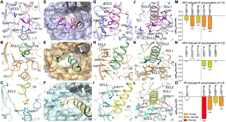
Fig. 3. Interactions between YR and the N terminus and α-helical region of the NPY peptide.
(A to C) Interactions between YR and the N-terminal residue Y/A1 of the NPY peptide. The salt bridges and hydrogen bonds are shown as red and green dashed lines, respectively. (D to F) Binding site for the N terminus of the NPY peptide in the YRs. The receptors are shown in both cartoon and surface representations. (G to L) Interactions between the NPY peptide and the extracellular loops of the YRs. (A, D, G, and J) NPY-Y1R-Gi1; (B, E, H, and K) NPY-Y2R-Gi1; (C, F, I, and L) PP-Y4R-Gi1. (M to O) NPY/PP-induced IP accumulation of YRs. (M) Y1R mutants; (N) Y2R mutants; (O) Y4R mutants. Bars represent differences in calculated peptide potency (pEC50) for each mutant relative to the wild-type (WT) receptor. Data are colored according to the extent of effect (EC50 ratio). Data are means ± SEM from at least three independent experiments performed in technical triplicate. *P < 0.01; ***P < 0.0001 by one-way analysis of variance followed by Dunnett’s post-test, compared with the response of the WT receptor. See table S1 for detailed statistical evaluation and expression level. The mutants on the left side of the dashed line are for the receptor residues involved in interaction with the N-terminal residue Y1/A1 of the NPY peptide, while the mutants on the right side are for the residues from the receptor extracellular loops.

