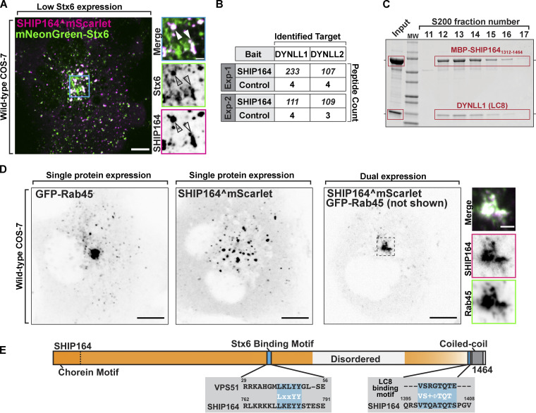
Figure 5.
Interaction of SHIP164 with proteins implicated in retrograde traffic to the Golgi complex. (A) Live fluorescence image of a COS-7 cell demonstrating partial colocalization of exogenous SHIP164^mScarlet (magenta) and mNG-Stx6 (green). Scale bar, 10 µm. High magnification of the indicated regions is shown at right, and the individual channels are shown as inverted grays. Arrowheads indicate overlapping fluorescence. Scale bar, 2 µm. (B) Mass spectrometry–based identification of material affinity-purified onto immobilized SHIP164 or onto a control protein from mouse brain extract. The selective enrichment of DYNLL1/2 peptides on the SHIP164 bait is shown. (C) A mixture of MBP-SHIP1641312–1464 and DYNLL1/2 was subjected to size exclusion chromatography. Coomassie Blue staining of SDS-PAGE of the eluted fractions reveals co-migration of the two proteins. (D) Live fluorescence images (inverted grays) of COS-7 cells expressing either GFP-Rab45 (left) or SHIP164^mScarlet (right), or both proteins together (only SHIP164 is shown) as indicated. Scale bar, 10 µm. High-magnification scale bar, 2 µm. (E) Cartoon of WT SHIP164 highlighting (blue) newly identified Stx6 and DYNLL1/2 interaction motifs (see also Fig. 1 A). Source data are available for this figure: SourceData F5.

