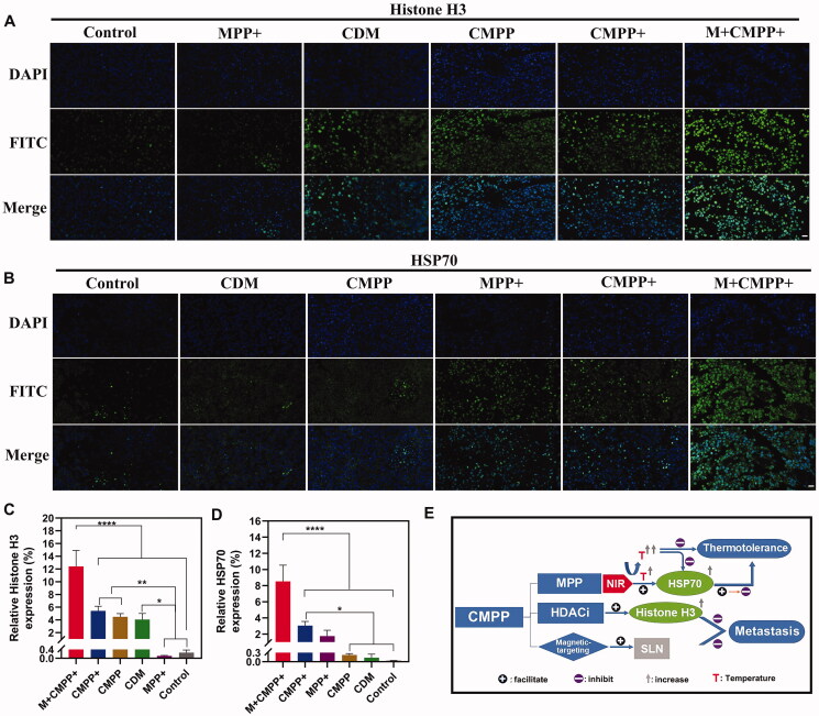
Figure 8.
Immunofluorescence staining analysis and flow chart of antitumor mechanism of CMPP. (A) Immunofluorescence staining analysis on Histone H3 (blue: cell nucleus; green: Histone H3) and (B) HSP70 (blue: cell nucleus; green: HSP70). Scar bar: 20 μm. The quantified expression levels of (C) Histone H3 and (D) HSP70 after immunofluorescence staining by image €(E) The antitumor mechanism of CMPP in vivo.

