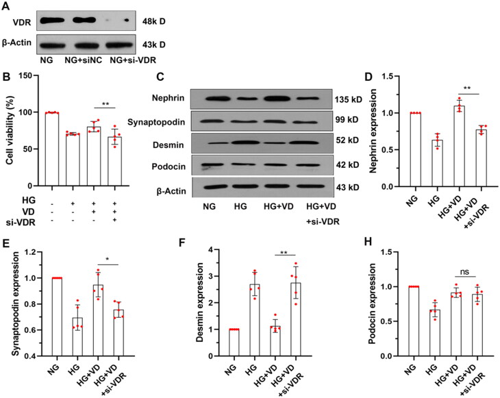
Figure 4.
VDR siRNA knocked down VDR reduced the protective effect of aVitD3 on MPC-5 injury Induced by high glucose. MPC-5 cells were transfected with VDR siRNA (si-VDR) or normal control siRNA (si-NC) and subjected to the indicated treatment. (A) Representative image of VDR immunoblot. (B) The MPC-5 cells' viability was determined by the CCK-8 assay. (C) Nephrin, Podocin, Synaptopodin and Desmin expression. Representative images from Western blot results. (D–H) Relative expression of proteins levels. Band densities were measured by the ImageJ program. The ratio of protein/actin density was calculated and normalized with the sham controls. Data were presented as mean ± SD. n = 5. **p < 0.01.

