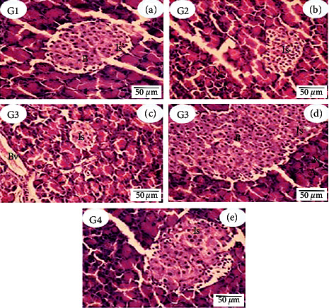
Figure 1.

Histopathological examination photomicrograph of the pancreatic section of control rats (G1, (a)) and Actinidia deliciosa (G2, (b)) groups showed the normal structure of islet of Langerhans cells (Is) gland, clumping of β-cells surrounded by small nuclei cells situated at the periphery of the gland, and acini gland (A) having epithelial cells with dark chromatic nuclei (N). Rats treated with STZ (diabetic rats) (G3, (c)) showed atrophy of islet (Is) Langerhans with reduction of the β-cells. The acinar cells stained strongly are arranged in lobules with prominent dark chromatin nuclei with marked dilation of blood vessels (Bv). (G3, (d)) Diabetic rat pancreas showed high proliferating β-cells (B), which crowded in the islet (appeared as small glands in structure). The large islet (Is) is surrounded by acini gland (A) with normal architecture epithelial cells with dark chromatic nuclei (N). The pancreas of diabetic rats that received Actinidia deliciosa (G4, (e)) showed normal proportions of the islet of Langerhans cells (Is) with mild edema (E) (H&E stain, bar = 50 µm).
