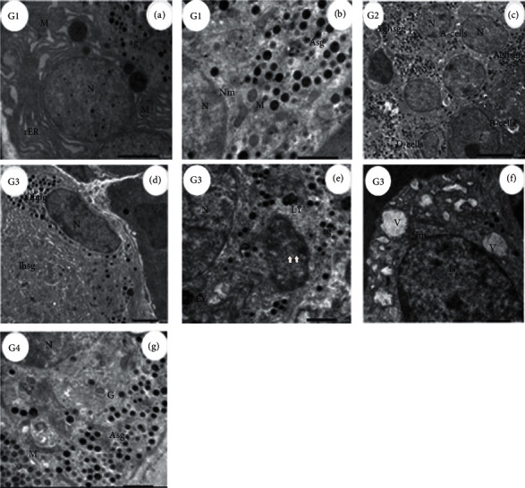
Figure 3.

Electron micrograph of Alpha-cells (α-cells) in different groups. (G1, (a)) Electron micrograph of rat pancreas showing a normal portion of exocrine with obvious nuclei (N) surrounded by rER containing zymogen granules (Z), the section showing also part of the endocrine (islets of Langerhans) revealing α-cells secretory granules (sg) and mitochondria (M). Scale bar = 2.0 µm, ×4000. (G1, (b)) Higher power view of control (G1) α-cells illustrating part of nuclei (N) with double nuclear membrane (Nm) and mitochondria (M) plus glucagon hormone-secreting granules (Asg). Scale bar = 1.0 µm, ×8000. (G2, (c)) ADAE-administered rats islets of Langerhans show two A-cells with nucleus (N) and normal glucagon hormone secretory granules (Aghsgr). The section showing also B-cells and D-cell (scale bar = 5.0 µm, ×1500). (G3, (d)) Diabetic rats α-cells with enlarged nuclei (N) and glucagon hormone-producing (Ghpg) + very few β-cells of insulin hormone-secreting granules (Ihsg) without a nucleus (scale bar = 2.0 µm, ×2500). (G3, (e)) High power view of α-cells in the pancreas of rats treated with STZ (G3) islets of Langerhans revealing part of the nucleus (N), cytoplasm with secretory granules (sg), autophagy (arrows), and lysosomes (LY) (scale bar = 1.0 µm, ×6000). (G3, (f)) Illustration of damaged α-cells, part of the nucleus (N), with double nuclear membrane (NM), absence of organoids in the cytoplasm, numerous vacuoles (V), and few secretory granules (sg) compared with control (scale bar = 1.0 µm, ×5000). (G4, (g)) Diabetic rat pancreas treated with ADAE revealing normal α-cells part of the nucleus (N) + cytoplasmic secretory granules (Asg), Golgi apparatus (G), and mitochondria (M) (scale bar = 1.0 µm, ×6000).
