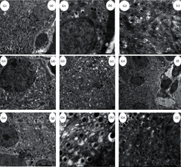
Figure 4.

Electron micrograph of Beta-cells (β-cells) in different groups. (G1, (a)) Control rat pancreas, islets of Langerhans denoting normal β-cells with nucleus (N), and normal secretory granules (Bsg) characterized by the electron-dense core and surrounded by a clear zone (cz) and area of A-cells secretory granules (Asg) and blood capillary (Bc) lined by large endothelial cell (En) with heterochromatic nuclei (HN) (scale bar = 2.0 µm, ×3000). (G1, (b)) Normal β-cells depicting the nucleus (N) (scale bar = 1.0 µm, ×8000). (G2, (c)) β-cells in islets of Langerhans of rat pancreas treated with ADAE showing part of the nucleus (N) with double nuclear membrane (NM), β-cells secretory granules (sg), Golgi apparatus (G) rER, and mitochondria (M) (scale bar = 1.0 µm. ×8000). (G3, (d)) Diabetic rat pancreas, β-cells with abnormal nuclei (N) with insulin hormone-secreting granules (Ihsg), mitochondria (M), and Golgi apparatus (G) (scale bar = 2.0 µm. ×2000). (G3, (e)) β-cells with pyknotic nuclei (PY) pyknosis + granules without secretion (gws) (scale bar = 2.0 µm. ×4000). (G3, (f)) β-cells with nuclei (N) with few granules (G) small in size and vacuolated rER + blood capillaries lined by 3 endothelial cells (En) (scale bar = 5.0 µm, ×1500). (G4, (g)) Islets of Langerhans of rat pancreas treated with STZ + ADAE denoting β-cells with segregated nuclear membrane (NM) and obvious nucleolus (Nu) and insulin-secreting granules with migrating granule apart of the cell (Bsg), rER, and mitochondria (M), glucagon hormone-secreting granules (Asg) without a nucleus, and ẟ-cells with somatostatin hormone-secreting granules (Dsg) apart of exocrine and rER (scale bar = 2.0 µm, ×3000. (G4, (h) and (i)) β-cells of diabetic rat + ADAE showing part of the nucleus (N), Golgi vesicles (Gv), rER, and insulin hormone-secreting granules (Bsg) in the cytoplasm) (scale bar = 500.0 nm, ×15000; scale bar = 1.0 µm, ×6000).
