
Figure 1.
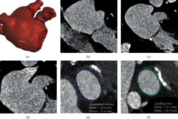
Anatomical structures and landmarks identified by the model. (a) 3D model reconstructed from the segmentation of the left atrium and left atrial appendage, where the landmarks of the anatomical ostium (blue), landing zone (green), fossa ovalis (yellow), and mitral annulus (red) are reported. (b) Fossa ovalis region indicated on the DICOM (yellow). (c) Mitral annulus indicated on the DICOM (red). (d) Anatomical ostium and landing zone indicated on the DICOM (blue and green, respectively). (e) Anatomical ostium plane. (f) Landing zone plane (Amulet device).