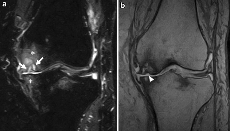
Fig. 14.
A 72-year-old female patient with knee OA. The patient complained of knee pain lasting for more than a year. a A coronal FS-T2WI shows severe cartilage damage and bony spur formation in the left medial femorotibial joint. Multiple subchondral cysts (arrows) and extended bone marrow edema-like signal intensity (asterisks) can be observed in the medial femoral and medial tibial condyles. b A coronal T2*-weighted image shows a slight deformity of the subchondral bone in the medial femoral condyle (arrowhead). This patient had been diagnosed with knee OA based on the clinical course, but the relatively old SIFK lesion was also a differential diagnosis

