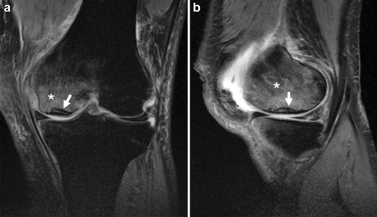
Fig. 4.
MRI of a 50-year-old man with a complaint of sudden left knee pain. a Coronal fat-suppressed proton density-weighted image (FS-PDWI), and b sagittal FS-PDWI show extensive bone marrow edema-like signal intensity over the medial femoral condyle (asterisk). A subchondral hypointense line is observed a few millimeters above the subchondral plate (arrow). Note that the line is almost parallel to the subchondral plate and open-ended in the medial part

