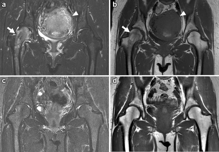
Fig. 8.
Coronal FS-PDWI and T1WI of a 40-year-old female patient in the postpartum period with transient BMES in the right hip. MRI was performed on the 12th day post-cesarean section for the complaint of severe right hip pain. a, b Initial MRI shows a diffuse bone marrow edema-like lesion extending from the right femoral head to the trochanteric region (arrow). No other obvious abnormal findings are noted, and the postpartum uterus is still enlarged (arrowhead). c, d The patient was treated conservatively, and her symptoms improved. A follow-up MRI taken 4 months later shows the disappearance of the bone marrow edema-like lesion in the right hip

