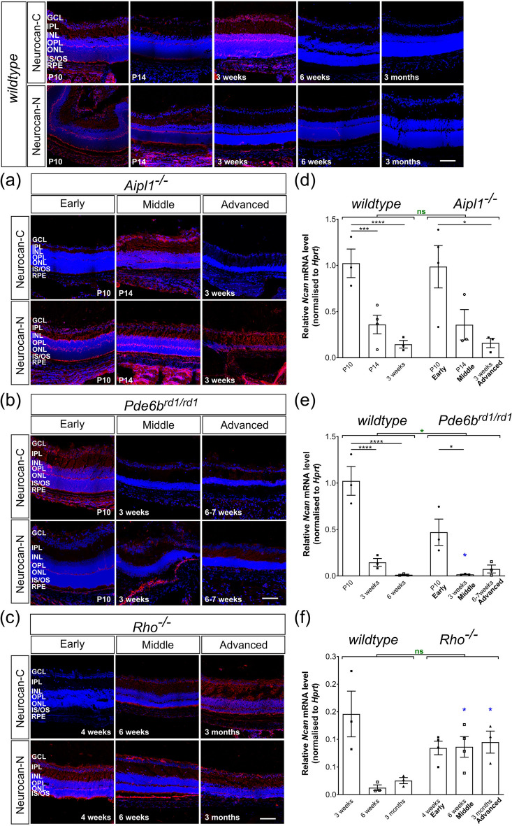
Figure 5.
Neurocan expression decreases with time and is unaffected by degeneration. Neurocan immunolabelling (red) was distributed throughout all the layers of the retina and low levels of mRNA transcripts were observed in adult retinae in all models. mRNA levels shown for wildtype are the same as in Fig. 1d. (a) (b) Immunolabelling of the Neurocan C-terminal fraction typically decreased in intensity in Aipl1-/- and Pde6brd1/rd1 mice with time, while that of the Neurocan N-terminal fraction remained constant in these models. (d) (e) mRNA levels of Ncan decreased with time in Aipl1-/-, Pde6brd1/rd1 and wildtype mice. (f) Ncan mRNA and (c) immunolabelling for both Neurocan-C and -N were largely unchanged across degeneration in Rho-/- mice. Scale bar, 100 µm. *p < 0.05, **p < 0.01, ***p < 0.001, ****p < 0.0001 (one-way ANOVA test with Bonferroni’s correction, black; unpaired t-test for age-matched comparisons between wildtype and disease model, blue; two-way ANOVA test applied for assessments of change over time, red). ONL—outer nuclear layer; OPL—outer plexiform layer; INL—inner nuclear layer; IPL—inner plexiform layer; GCL—ganglion cell layer. Nuclei are counter stained with Dapi (blue).

