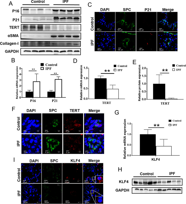
Fig. 1. KLF4 and TERT expression was downregulated in AECs in human IPF lung tissues.
A Western blotting was performed to evaluate the expression of P16, P21, TERT, αSMA and Collagen-I in IPF lung tissue and control lung tissues. Four randomly selected sample from each group were shown. B, D qPCR were performed to test P16, P21 and TERT in human IPF lung tissues and control group. C Double-labeled immunofluorescent staining were performed to examine the expression of SP-C (green) and P21 (red) in AECs. E Quantification of the protein level of TERT. F Double-labeled immunofluorescent staining were performed to examine the expression of SP-C (green) and TERT (red) in AECs. G, H qPCR and western blotting were performed to test KLF4 expression level in human IPF lung tissues and control group. I Immunofluorescent staining were performed to examine the expression of SP-C (green) and KLF4 (red) in AECs. *P < 0.05, **P < 0.01, ***P < 0.001 by t-test.

