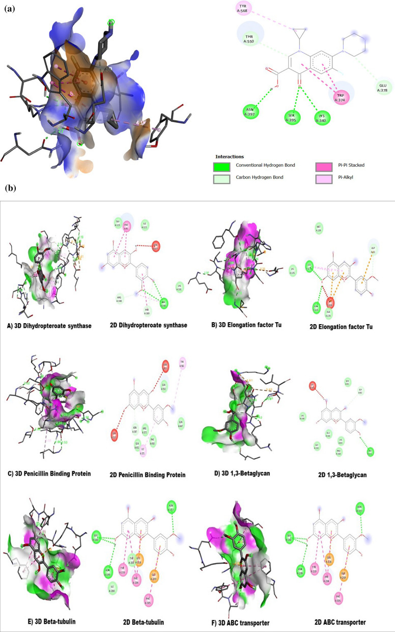
Figure 6.
(a) The 2D and 3D intermolecular contact between ciprofloxacin and Pencillin binding protein. Chemical structures were drawn by ChemDraw Pro 16.0 Suite (PerkinElmer, USA) and analyzed by the Discovery studio visualizer (BIOVIA Discovery studio 2020 Client). (b) The 3D and 2D intermolecular contact between A) Dihydropteroate synthase B) Elongation factor Tu and C) Penicillin Binding Protein D) 1,3-Betaglycan E) Beta-tubulin F) ABC transporter with peonidin . Chemical structures were drawn by ChemDraw Pro 16.0 Suite (PerkinElmer, USA) and analyzed by the Discovery studio visualizer (BIOVIA Discovery studio 2020 Client).

