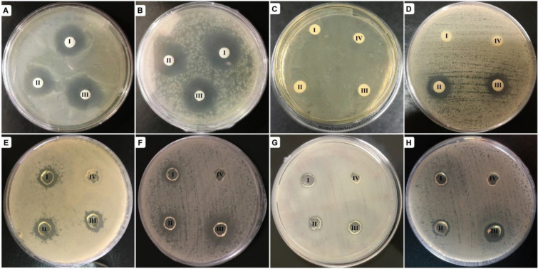
Figure 5.
Inhibition zones around the prepared hydrogels. (A–D) Non-cross-linked and (E–H) cross-linked hydrogels. The plates in (A), (C), (E), and (G) are Escherichia coli cultures. The plates in (B), (D), (F), and (H) present Staphylococcus aureus cultures. In each plate, the hydrogels numbered as I–III contain 0.03, 0.08, and 0.15 mM nisin; number IV represents the control sample. In the plates displayed in (A) and (B), the controls are avoided due to repetition in the other 6 plates.

