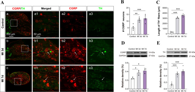
Fig. 2.
Sprouting of sympathetic post-ganglionic fibers in DRGs after MI. A Representative images of immunoreactivity to calcitonin gene-related peptide (CGRP, red, a marker for peptidergic nociceptive neurons) and tyrosine hydroxylase (TH, green) which labels adrenergic fibers in the DRGs of control and MI rats at 3 and 7 days after LAD ligation. a1–3: the higher power views of the boxed area in a (the same for b and c). White arrows indicate sprouted sympathetic terminals. B, C Quantification of the number of CGRP+ neurons and the length of TH+ fibers in the DRGs (n = 4/group, **P <0.01, ***P <0.001). D, E Western blots showing increased levels of CGRP and TH protein expression in the C8–T6 DRGs in rats at days 3 and 7 post-MI (n = 3–4/group, *P <0.05, ***P <0.001). One-way ANOVA with least significant difference multiple comparisons in B–E.

