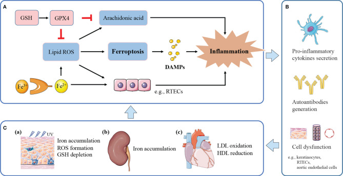
Figure 2.
The potential model of ferroptosis in lupus inflammation and manifestations. (A) Ferroptosis releases DAMPs to trigger inflammation. Iron and ROS accumulation promote a pro-inflammatory environment. Massive lipid ROS released by ferroptosis helps to convert arachidonic acid to inflammatory mediators. GPX4 suppresses inflammation by inhibiting arachidonic acid oxidation and lipid peroxidation. (B) Ferroptotic cell death and induced inflammation exert causative effects in SLE through pro-inflammatory cytokines secretion, and autoantibodies generation, finally leading to cell dysfunction and tissue damage. (C) (a) Inflammation induced by UV irradiation amplifies inflammatory and immune responses, eventually causing cutaneous lesions. UVB-exposed skin lesions exhibit iron accumulation, excessive ROS and GSH depletion, leading to keratinocytes ferroptosis. (b) Persistent inflammation and immune complexes deposition accelerate lupus progression to renal failure. Kidneys uptake excessive iron in the renal tubules and undergo ferroptosis under pathological conditions. (c) Lipid peroxidation and induced inflammation contribute to endothelial dysfunction and cardiovascular injury. Lupus patients with progressive atherosclerosis show decreased HDL and increased oxLDL, which may further promote ferroptosis in aortic endothelial cells.

