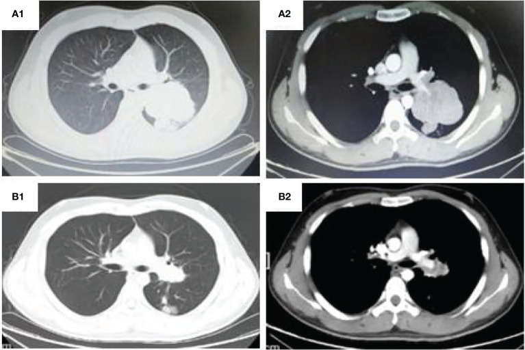
Figure 1.
CT thorax: (A1, 2) (Shanxi Provincial Cancer Hospital,2021-02-03) shows a large irregular and well-defined border mass lesion measuring 7.7×6.3 cm with inhomogenous enhancement beside the left hilus pulmonis across the interlobar fissure and an enlarged mediastinal lymph node measuring 2.7×1.9 cm. (B1, 2). (Shanxi Provincial Cancer Hospital,2021-05-12)Tumor was remarkable reduced to 2.5*3.4cm and the mediastinal lymph node was 1.2 cm.

