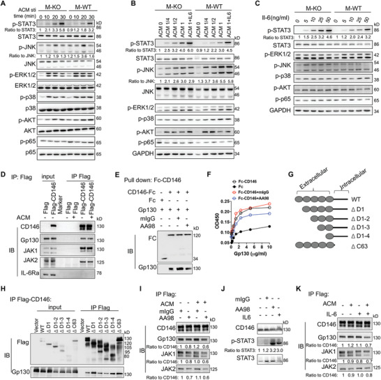
Figure 6.

CD146 interacts with Gp130 and negatively regulates the STAT3 activation. Immunoblot analyses of BMDMs stimulated A) with ACM for the indicated times, or B) at the indicated concentrations, or C) with IL‐6 at the indicated concentrations using antibodies specific to ERK, JNK, p38, AKT, STAT3, and p65 and/or their phosphorylated forms (representative of n = 3). The ratio indicates quantification of the phosphorylated form relative to total protein. D) Immunoblot analysis (IB) of CD146, Gp130, JAK1, JAK2, and IL‐6Rα in RAW264.7 cells transfected with CD146‐Flag plasmid or control Flag plasmid and stimulated with ACM followed by immunoprecipitation (IP) with Flag‐beads (representative of n = 3). E) Pull‐down assay of CD146‐Fc and Gp130 proteins in the presence of AA98 or control mouse IgG (mIgG) (representative of n = 3). F) Fc or Fc‐CD146+ with mIgG or AA98 was added to wells coated with different concentrations of Gp130, and ELISA was performed (representative of n = 3). G) Schematic representations of recombinant CD146 extracellular domains. H) Immunoblot analysis (IB) of Flag and Gp130 in 293T cells transfected with CD146‐Flag plasmid followed by IP with Flag‐beads (representative of n = 3). I) Immunoblot analysis (IB) of CD146, Gp130, JAK1, and JAK2 in RAW264.7 cells transfected with CD146‐Flag plasmid (RAW264.7‐CD146‐Flag) and treated with or without ACM plus mIgG or AA98 (50 µg mL−1) for 12 h (representative of n = 3). Ratios indicate quantification of the protein levels relative to CD146. Data are representative of at least three independent experiments. J) IB of p‐STAT3 in RAW264.7‐CD146‐Flag treated with or without ACM plus IL‐6 (100 ng mL−1), mIgG, or AA98 (50 µg mL−1) for 10 min (representative of n = 3). Ratios indicate quantification of the p‐STAT3 protein levels relative to STAT3. CD146 served as control. K) IB of CD146, Gp130, JAK1, and JAK2 in RAW264.7 cells transfected with CD146‐Flag plasmid and treated with or without ACM plus IL‐6 (100 ng mL−1) for 12 h (representative of n = 3). Ratios indicate quantification of the protein levels relative to CD146.
