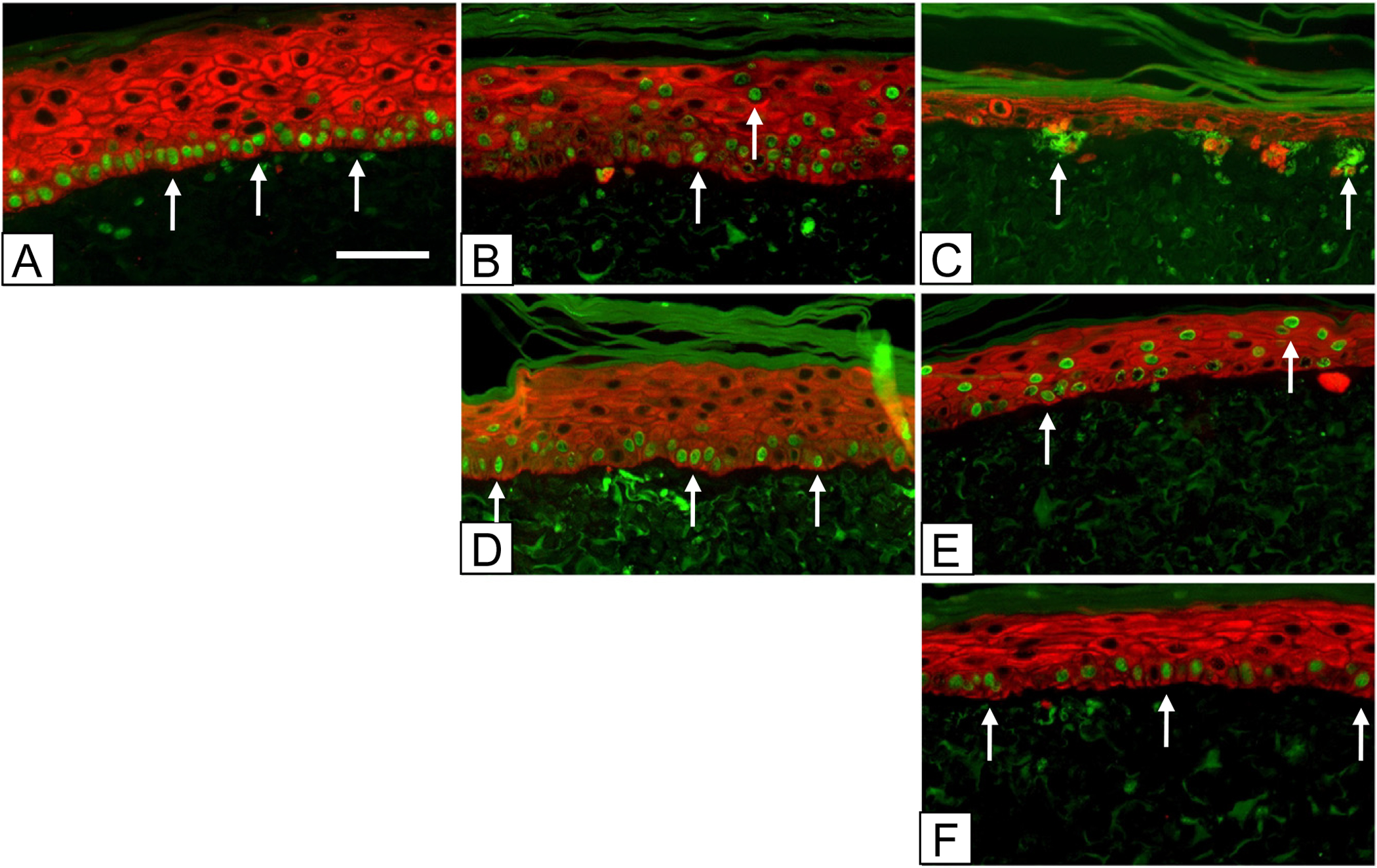
Figure 1.

Immediate and serial chases over 3 weeks in vitro. Nuclei in ESS were stained green with anti-BrdU-FITC at incubation days: (A–C) 5–7 (top row); (D, E) 12–14 (center row); and (F) 19–21 (bottom row). Keratinocytes were stained red with Alexafluor 594 as described in the Methods. Samples were prepared for fluorescence immunohistochemistry at incubation days: (A) 8 (left column); (B, D) 15 (center column); and (C, E, F) 22 (right column). The time course shows a progressive decrease in numbers of labeled nuclei and reorganization of label-retaining keratinocytes into clusters at the basement membrane (arrows). Scale bar = 0.1 mm.
