
Figure 1.
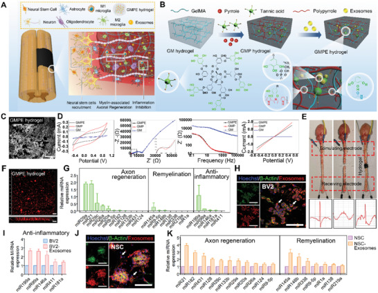
Characteristics of the GMPE hydrogels. A) Illustration of how the GMPE hydrogel can reduce early inflammation, enhance NSCs recruitment and promote myelin‐associated axonal regrowth to synergistically promote locomotor recovery after spinal cord hemisection. B) The three‐step synthesis procedure for the GMPE hydrogel was illustrated. The GMP hydrogel was synthesized by TA interacting with the amide bond on the GM backbone and the nitrogen groups on PPy chains. BMSC‐exosomes were reversibly immobilized into GMP hydrogels via hydrogen bond formation between TA polyphenol groups and phosphate groups in exosomes phospholipid to form GMPE hydrogel. C) Microstructure of the GMPE hydrogel was observed by SEM. Scale bars: 25 µm. D) Electrical characterization, including CV, EIS, I–V, and Bode plots of GMP and GMPE hydrogels showed excellent electrical performance. E) After the isolation of transected spinal cords, the stimulating electrical signals were retransmitted by GMPE hydrogels. F) IF imaging showed that exosomes were evenly distributed into the GMPE hydrogel and the penetration depth of the exosomes was more than 100 µm. Scale bars: 100 µm. G) RT‐qPCR indicated that BMSC‐exosomes express of axonal regeneration‐related, remyelination‐related, and anti‐inflammatory‐related miRNAs (n = 3). H) BV2 cells cultured on the GMPE hydrogel can normally phagocytize exosomes released from the hydrogel. White arrows indicate where BV2 cells have phagocytosed exosomes. Scale bars: 100 µm. I) Anti‐inflammatory‐related miRNAs expression increased as the result of BV2 cells phagocytosing exosomes (n = 3). J) PKH26‐labeled exosomes were clearly detected in the cytoplasm of NSCs, suggesting successful in vitro endocytosis of exosomes released from the GMPE hydrogel. White arrows indicate where NSCs have phagocytosed exosomes. Scale bars: 100 µm. K) Axonal regeneration‐related and remyelination‐related miRNAs expression increased after NSCs phagocytize exosomes (n = 3).