
Figure 4.
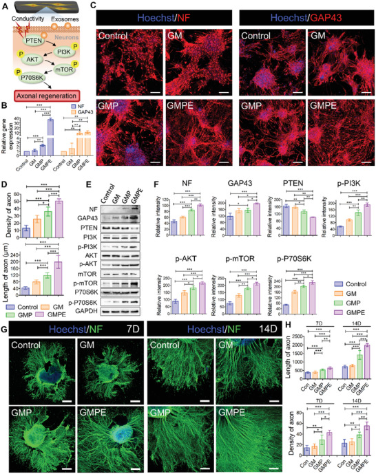
Axon outgrowth on hydrogels. A) Illustration of the BMSC‐exosomes electroconductive hydrogel mechanism and synergistic promotion of axon outgrowth through the activation of the PTEN/PI3K/AKT/mTOR pathway. B) RT‐qPCR indicating that GMPE hydrogels can promote NF and GAP43 gene expression (n = 3). C) IF images of the axon‐associated proteins NF and GAP43 in NSCs grown on hydrogels for 7 days. Red IF represents the NF or GAP43, respectively. Scale bars: 100 µm. D) The density (n = 5) and length (n = 11) of axons were quantified using ImageJ software. E) WB result of the expression of NF and GAP43 proteins and the relative protein expression of the PTEN/PI3K/AKT/mTOR pathway in NSCs cultured on control or hydrogel conditions for 7 days. F) Protein band intensity was quantified (n = 3). G) IF images of the NF positive axon in DRGs grown on hydrogels for 7 days. H) The density (n = 5) and length (n = 11) of axons were quantified using ImageJ software. Statistical differences were determined using an ANOVA with Bonferroni's multiple comparison test (* p < 0.05, ** p < 0.01, *** p < 0.001).