
Figure 4.
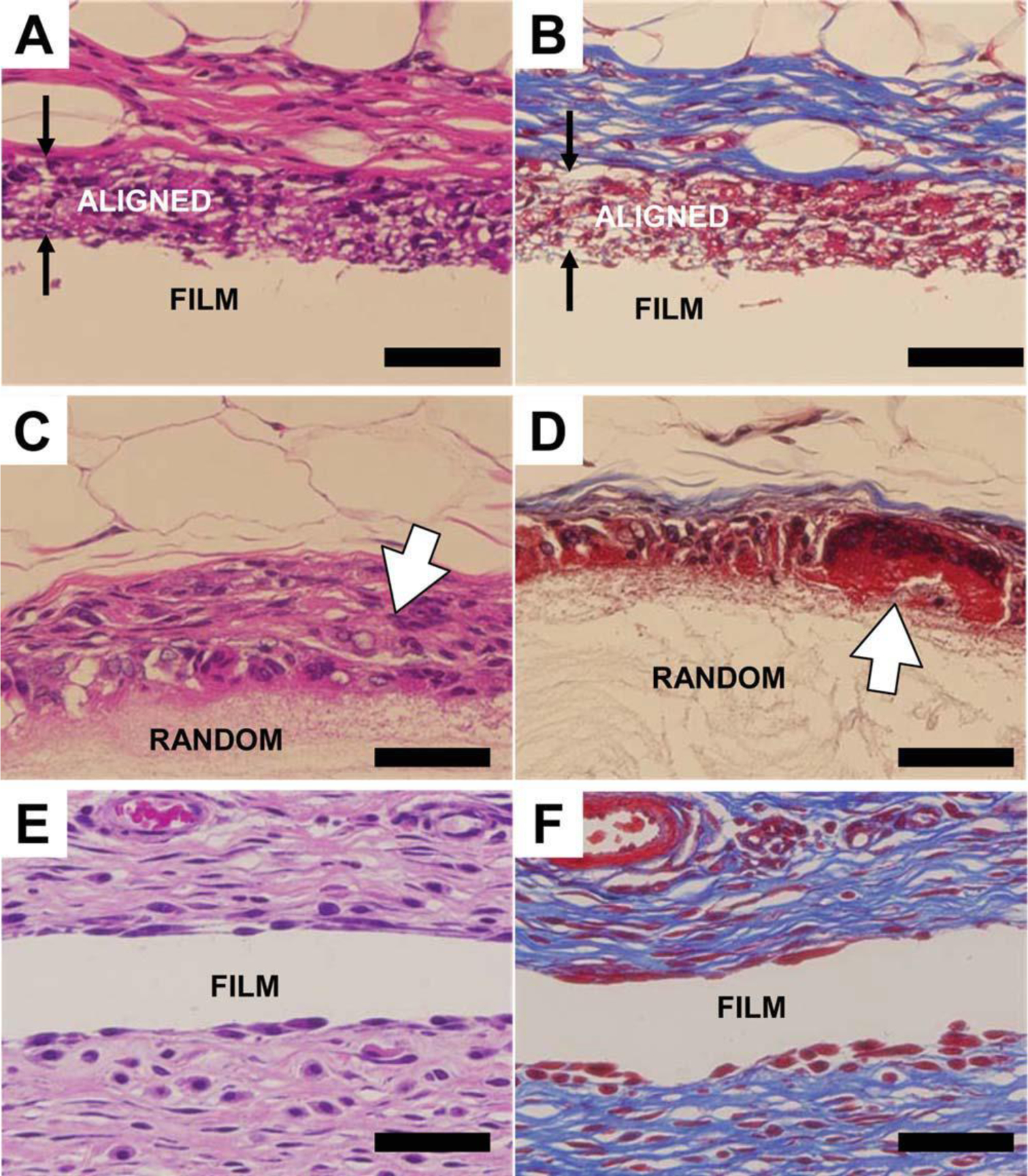
Histological images showing the fibrous capsules formed on PCL scaffold surfaces at week 4. (A,B) Aligned fibers. (C,D) Random fibers. (E,F) Film. Left, H and E staining. Right, Masson’s Trichrome staining. Three types of scaffolds are indicated on the images. The paired arrows indicate the aligned scaffold with cell infiltration. The open arrows show the strong macrophage/foreign body giant cell reaction on the random fiber scaffold. The scale bar is 50 µm.