
Figure 5.
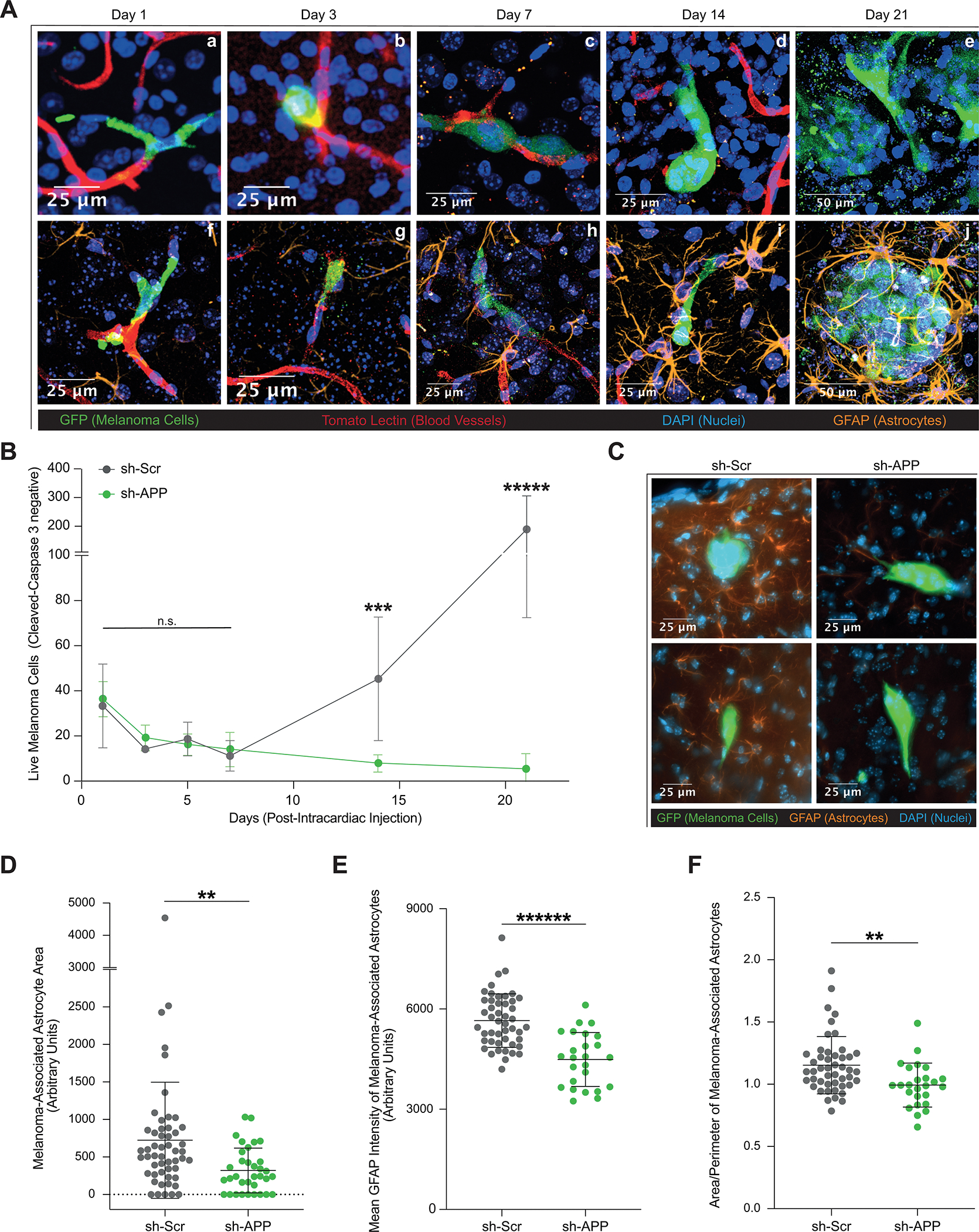
Melanoma-secreted Aβ is Required for Growth and Survival in the Brain Parenchyma. A, Representative images of brain slice immunofluorescence of 12–273 BM cells post intracardiac injection in NSG mice at days 1(Aa,Af), 3 (Ab,Ag), 7(Ac,Ah), 14 (Ad,Ai), and 21 (Ae,Aj). Fluorescent markers: green = anti-GFP (melanoma cells), red = tomato lectin (blood vessels), blue = DAPI (nuclei), orange = anti-GFAP (astrocytes). B, Quantification of live 12–273 BM cells in the brain parenchyma over time after intracardiac injection (6 mice per group per experiment, 4 50uM-thick brain slices per mouse). Day 14 sh-Scr vs. sh-APP (*** p< 0.0005), day 21 sh-Scr vs. sh-APP (***** p<0.000005). C, Representative images of brain slice immunofluorescence at day 10 post intracardiac injection. D-F, Two-dimensional quantification of parameters of astrocyte reactivity in sh-Scr vs. sh-APP melanoma-associated astrocytes (2 mice per group, 3 50uM-thick brain slices per mouse). D, Area. sh-Scr vs. sh-APP (** p<0.005). E, Mean GFAP Intensity. sh-Scr vs. sh-APP (****** p<0.0000005). F, Area/Perimeter – a proxy measurement for hypertrophy. sh-Scr vs. sh-APP (** p<0.005).