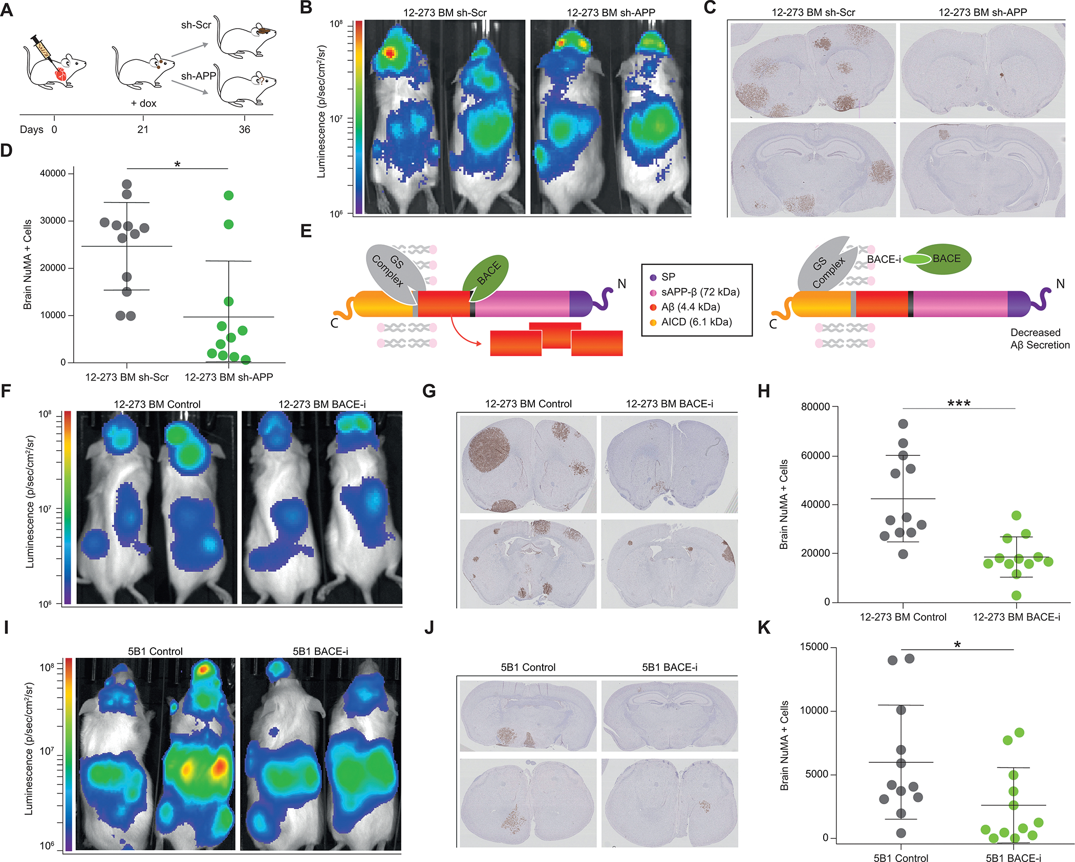
Figure 7.

Aβ is a Promising Therapeutic Target for Treatment of Brain Metastasis. A, Diagram of therapeutic simulation experiment inducing silencing of APP in established brain metastases. B-D, Induction of silencing of APP in established brain metastases (11–12 NSG mice per group). B, Representative IVIS images at day 37 post intracardiac injection. C, Representative images of FFPE brain slides with labeling of metastatic cells by anti-NuMA immunohistochemistry. D, Quantification of NuMA+ metastatic cells in mouse brains. sh-Scr vs. sh-APP (* p<0.05). E, Diagram of APP cleavage and beta-secretase inhibition of Aβ production. F, Representative IVIS images at Day 28 post intracardiac injection with 12–273 BM STC (12 NSG mice per group). G, Representative images of FFPE brain slides with 12–273 BM metastatic cells stained by anti-NuMA immunohistochemistry. H, Quantification of NuMA+ metastatic cells in mouse brains. 12–273 BM Control vs BACE-i (*** p<0.0005). I, Representative IVIS images at Day 28 post intracardiac injection with 5B1 melanoma cell line (12 NSG mice per group). J, Representative images of FFPE brain slides with 5B1 metastatic cells stained by anti-NuMA immunohistochemistry. K, Quantification of NuMA+ metastatic cells in mouse brains. 5B1 Control vs BACE-i (* p<0.05).
