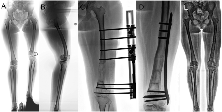
Figure 20.
(A) Radiographic images of both legs (when standing) of a 20-year-old girl with distal femoral fracture sequelae. A 9-cm left femur shortening and 15˚ distal femur varus deformity on the frontal plane were observed. (B) Lateral one-leg standing radiography shows a 20˚ distal femur procurvatum deformity. (C) Acute distal axis correction and gradual lengthening with a monolateral external fixator was performed. (D) After achieving length correction, percutaneous plating. (E) Radiographic images of both legs standing after 6 months of hardware removal.

 This work is licensed under a