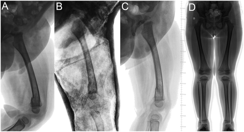
Figure 13.
(A) Lateral radiographic image of the right femur of a 2-year-old boy, with a distal femoral fracture. (B) Radiographic control at 1 week. (C) Radiographic image after cast removal at 4 weeks. (D) Radiographic images (anteroposterior view) of both legs when standing, after 1 year, with no limb-length discrepancy or axis deviation.

 This work is licensed under a