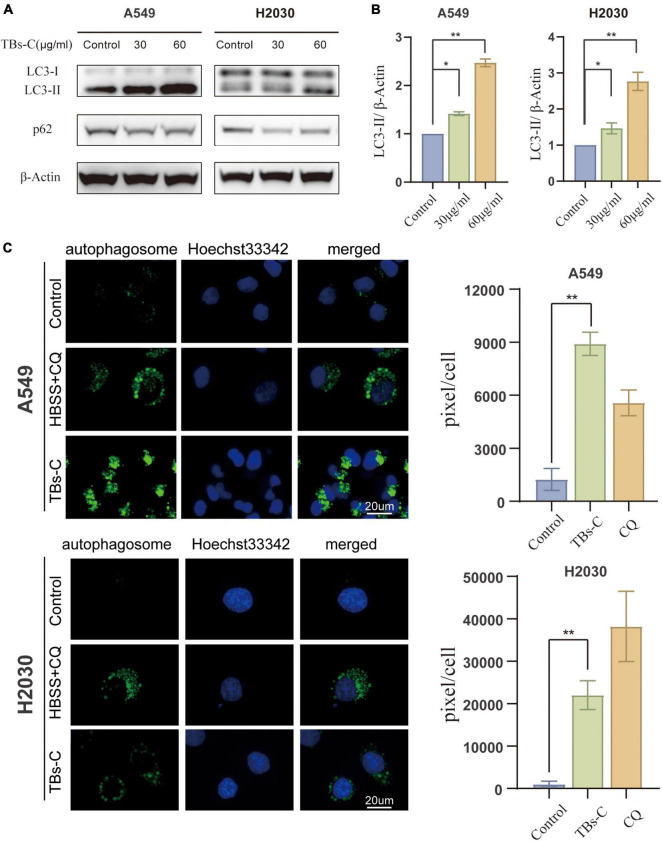
FIGURE 3.
Accumulation of autophagosomes in TBs-C treated lung cancer cells. (A) Western blotting results of the LC3 and p62 protein expression in TBs-C treated and untreated cells. (B) The densitometric analysis of the changes in the abundance of LC3-II normalized to the β-actin level. (C) The indicated cells were treated with TBs-C (60 μg/mL) or untreated for 24 h. The cells were labeled with CYTO-ID® green detection reagent at the end of the treatment period to detect the autophagosomes. As a positive control for autophagosome accumulation, the cells were treated with HBSS in the presence of CQ (60 M) for 3 h. The puncta areas per cell are given in three randomly selected images for each culture condition, *p < 0.05, **p < 0.01.

