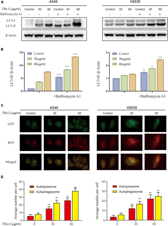
FIGURE 4.
Treatment with TBs-C promotes the autophagic flux in NSCLC cells. (A) After 24 h of treatment with different concentrations of TBs-C, the cells were treated with or without baf A1 (100 nmol/mL for 3 h), and the LC3 expression was evaluated by Western blotting. (B) Densitometric analysis of the changes in LC3-II abundance normalized to the β-actin level. (C,D) The indicated cells were transfected with the RFP-GFP-LC3B Premo™ Autophagy Tandem Sensor. Fluorescence microscopy was used to count the red and yellow puncta (Scale bar = 20 m). The number of yellow and red puncta in each cell was calculated from at least 20 cells from each group, *p < 0.05, **p < 0.01.

