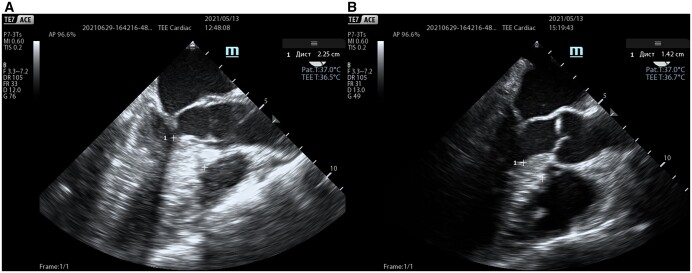
Figure 2:
Intraoperative TEE images (mid-oesophageal aortic valve long-axis view) in early systole before (A) and after (B) procedure; elimination of the left ventricular outflow tract obstruction with concomitant procedures on the mitral valve subvalvular apparatus. Septal thickness (1) was measured before and after correction. TEE: transoesophageal echocardiography.

