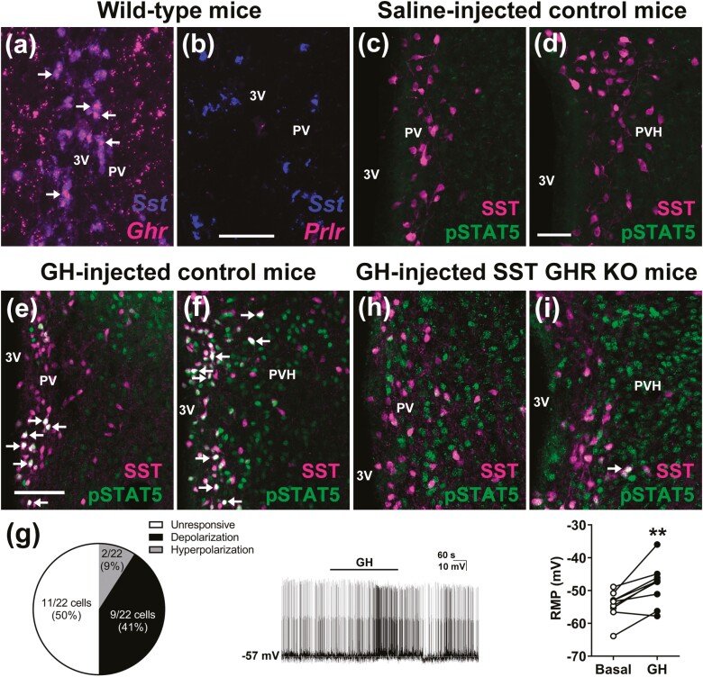
Figure 1.
PV/PVHSST neurons express Ghr mRNA and are responsive to GH. (A,B) Colocalization between Sst mRNA and Grh mRNA, and Sst mRNA and Prlr mRNA in the PV of mice. Scale bar = 100 µm. Abbreviations: 3V, third ventricle. The arrows indicate double-labeled neurons. (C,D) PV/PVHSST neurons exhibit no STAT5 phosphorylation (pSTAT5) in saline-injected mice. Scale bar = 50 µm. (E,F) PV/PVHSST neurons exhibit pSTAT5 after an IP GH injection. (G) Electrophysiological recordings of PV/PVHSST neurons after GH application (22 neurons from 8 mice). Representative recording of a cell that showed significant depolarization a few minutes after GH application to the bath. Changes in the resting membrane potential (RMP) of depolarized neurons (n = 9; **P < .01; paired t-test). (H,I) SST GHR KO mice show only few GH-induced pSTAT5 in PV/PVHSST neurons, demonstrating the efficacy of the cell-specific GHR ablation.

