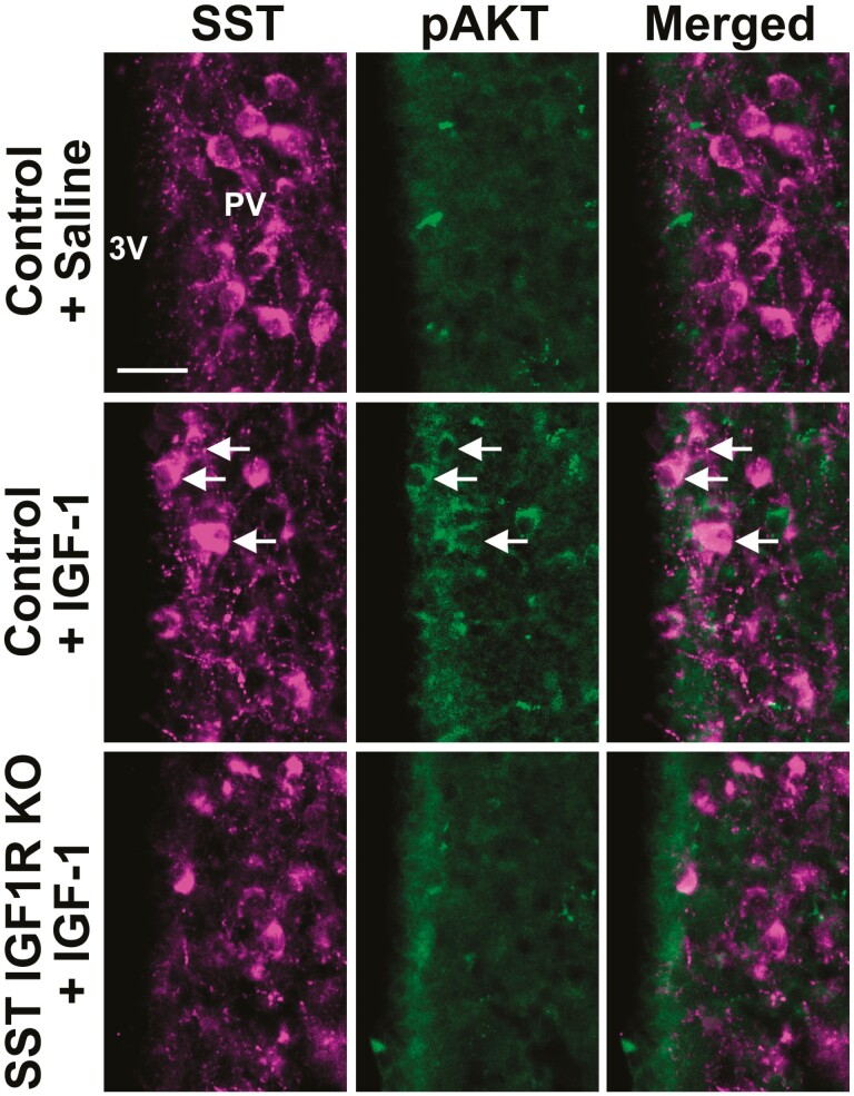
Figure 6.
IGF-1 induces AKT phosphorylation in PVSST neurons of control mice but not in SST IGF1R KO mice. Fluorescence photomicrographs showing the colocalization between SST and pAKT in the PV of control mice that received ICV injection of saline or IGF-1, and in SST IGF1R KO mice that received ICV IGF-1 injection. The arrows indicate double-labeled neurons. Abbreviation: 3V, third ventricle. Scale bar = 25 µm.

