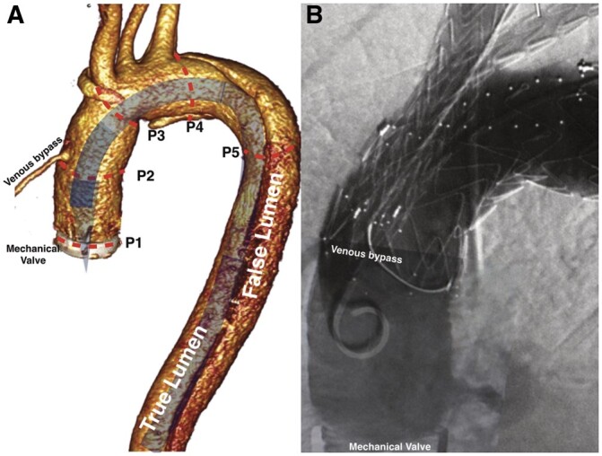
Figure 1:

(A) 3-Dimensional reconstruction of the tomographic image. Expected position of a delivery system without tip modification. Sizing: P1–P2: space for the tip of the delivery system (29 mm); P2-P3: proximal sealing zone (34 mm); P3-P4: access to the branches (50 mm); P4-P5: distal sealing zone. (B) Completion angiographic scan: patent venous bypass; no endoleak.
