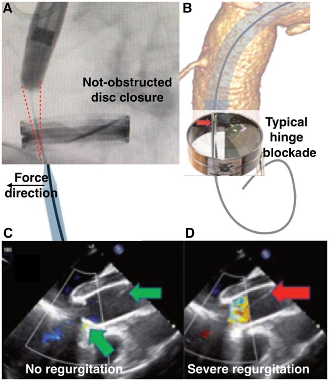
Figure 2:

(A) Red sketch on the fluoroscopic image shows space obtained after cutting 15 mm from the tip of the stent graft. The wire was guided by the 6 Fr sheath from the apex to keep it away from the disc hinge. (B) 3-Dimensional reconstruction of the tomographic image. Mechanism of the disc blockade if the valve is simply crossed. (C/D) Transoesophageal echocardiography. (C) Effective valve closure; green arrows show correct position of the wire. (D) Severe valve regurgitation after the blockade of the disc. Red arrow shows the position of the wire.
