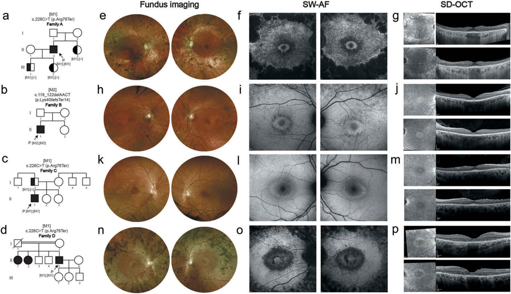
Figure 1.
Early parafoveal RPE loss in four individuals affected with RP.
(a-d) Pedigrees of family A-D are demonstrated. Equal signs denote the wild-type allele, square boxes indicate males, circles indicate females, and affected individuals are pointed out in black. Arrows indicate the probands. Double-line indicates consanguineous marriages. M1, and M2 depict the positions of the mutations identified in this study. (e-g) Images of both eyes of A-II:2 at 62-year-old. Fundus imaging (e) shows attenuated vessels, waxy pallor of the optic disc, atrophic changes and bone spicule pigmentation in the midperiphery area. SW-AF (f) reveals a hyperautofluorescent ring surrounding the central area of the macula with surrounding parafoveal and mid-peripheral severe flaky or fused hypoautofluorescence. SD-OCT (g) reveals outer retinal atrophy with centrally preserved RPE and ellipsoid zone (EZ) line, with hypertransmission outside the corresponding area of choroid. (h-j) Images of both eyes of B-II-1 at 60-year-old. Fundus imaging (h), SW-AF (i), and SD-OCT (j) show moderate pigmentary changes in peripheral retina outside the vascular arcades with hyperautofluorescent ring around the macular, and surrounding parafoveal and mid-peripheral mottling hypoautofluorescence, as well as outer retinal atrophy with centrally preserved RPE and EZ line, with hypertransmission outside the corresponding area of choroid. (k-m) Images of both eyes of C-II:1 at 21-year-old. Fundus imaging (k), SW-AF (l), and SD-OCT (m) show mild pigmentary changes in peripheral retina outside the vascular arcades, and hyperautofluorescent ring around the macula with mild parafoveal loss of autofluorescence, as well as thinning of the outer retina with centrally preserved RPE and EZ line. (n-p) Images of both eyes of D-II:5 at 49-year-old. Fundus imaging (n) shows severe pigmentary changes with bone spicule pigmentation, as well as attenuated vessels, and the waxy pallor of the optic disc. Hyperautofluorescent ring surrounding the normal-looking macular and surrounding parafoveal and mid-peripheral severe flaky and mottling hypoautofluorescence are seen on SW-AF (o). SD-OCT images (p) reveal outer retinal atrophy with centrally preserved RPE and ellipsoid zone (EZ) line, with hypertransmission outside the corresponding area of choroid.

