Figure 6.
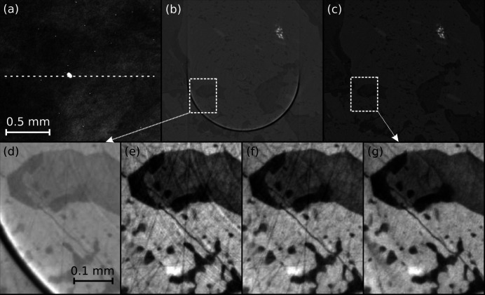
Reconstruction of parallel beam CT data with tofu ez. The sample is metamorphic schist (a piece of rock composed of four minerals). The top row shows from left to right fragments of: a raw CT projection (a) (dashed line indicating reconstructed row); of a slice reconstructed from phase-retrieved projections without the application of any artifact-reduction algorithms (b); of the same slice reconstructed with suppression of artifacts (c). Magnified fragments of images (b) and (c) are shown in insets (d) and (g), respectively. Images in the bottom row demonstrate progressive improvement when: only phase-retrieval was applied to data (d); phase-retrieval and broad ring removal (e); phase-retrieval, broad and narrow ring removal (f); all previous algorithms were applied and the outliers were removed from projections and flat-field images (g). Panels (a) and (d)–(g) have been inserted after automatic contrast adjustment was applied in ImageJ once to the entire image; no image correction of any kind was applied to panels (b, c). The scalebar for all images in a row is the same as shown in the heading image of that row.

