Figure 6.
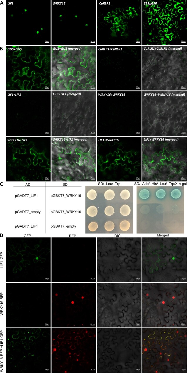
Subcellular localization of candidate genes and protein–protein interactions. A, Subcellular localizations of LIF1, SlWRKY16, and CuRLR1 proteins. B, Verification of protein–protein interactions and locations of SlWRKY16 and LIF1 by BiFC. The gene with cCitrine fusion is listed before the “+” sign and the gene with nCitrine fusion is listed after the “+” sign. C, Yeast two-hybrid (Y2H) results for interaction between LIF1 and SlWRKY16. The plasmids with GAL4 activation domain (AD) and GAL4 DNA binding domain (BD) were co-transformed to yeast AH109 competent cells. Transformed yeast cells were screened on SD/–Leu/–Trp medium plates to select successful co-transformants and then assayed by culturing on high-stringency SD/–Ade/–His/–Leu/–Trp medium plates with 40 μg·mL−1 X-α-Gal. The positive protein–protein interactions between LIF1 and SlWRKY16 are indicated by growth on SD/–Ade/–His/–Leu/–Trp/X-α-Gal medium plates and blue colony color. D, Co-expression of fusion protein LIF1-GFP and SlWRKY16-RFP to observe subcellular localizations. Yellow color in the merged panel indicates that GFP and RFP signals are overlapped. Scale bars for A, B, D: 20 µm.

