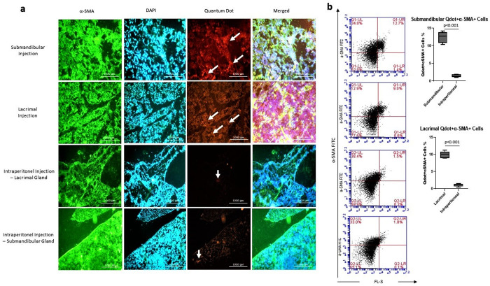
Fig 8. The differentiation analysis of DFMSCs into epithelial lineage in glandular tissues.
(a) The fluorescent microscope images of glandular tissues. Glandular tissues were stained with α-SMA (FITC) and DAPI, and analyzed for Quantum dot 655 (Qdot-655) labeled cells. Submandibular and lacrimal injections of DFMSCs showed high differentiation into glandular epithelial cells on day 28 compared to the intraperitoneally injected DFMSCs. (b) Flow cytometry analysis for α-SMA stained cells within Qdot-655 labeled cells. The submandibular and lacrimal injections of DFMSCs highly express α-SMA in submandibular and lacrimal glands of SS mouse model compared to intraperitoneal injections (p<0.001 and p<0.001, respectively).

