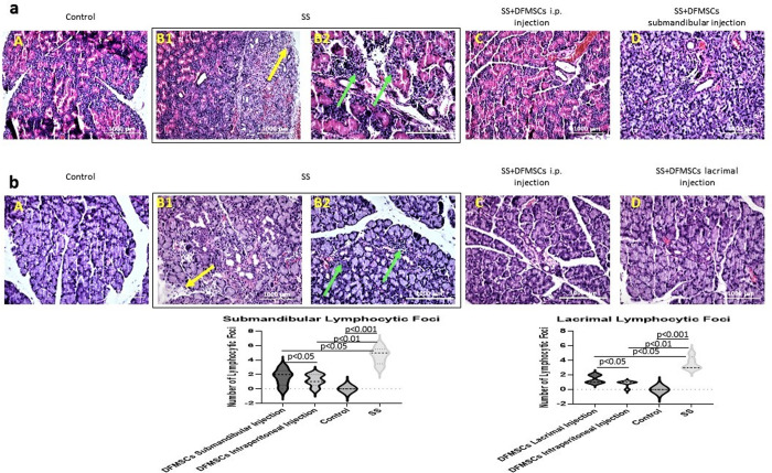
Fig 9. Histopathological analysis of glandular tissues.
(a) Hematoxylin-eosin staining of submandibular and (b) lacrimal glands. The statistical analysis of lymphocytic foci of glandular tissues. Submandibular and lacrimal glands of SS mice showed high amounts of lymphocytic foci (Focus score >1) compared to control subjects (Focus score <1) (p<0.001). The lymphocytic foci were shown with green arrows. Tissue destruction and fibrosis in submandibular and lacrimal glands in Group 2 were observed in the glandular sections (yellow arrows). DFMSCs significantly reduced lymphocytic infiltrates in submandibular glands (p<0.01) and in lacrimal glands (p<0.05), and ameliorated tissue destruction when applied intraperitoneally. Submandibular or lacrimal injection of DFMSCs significantly reduced lymphocytic infiltration and fibrosis in submandibular (p<0.05) or lacrimal glands (p<0.05).

