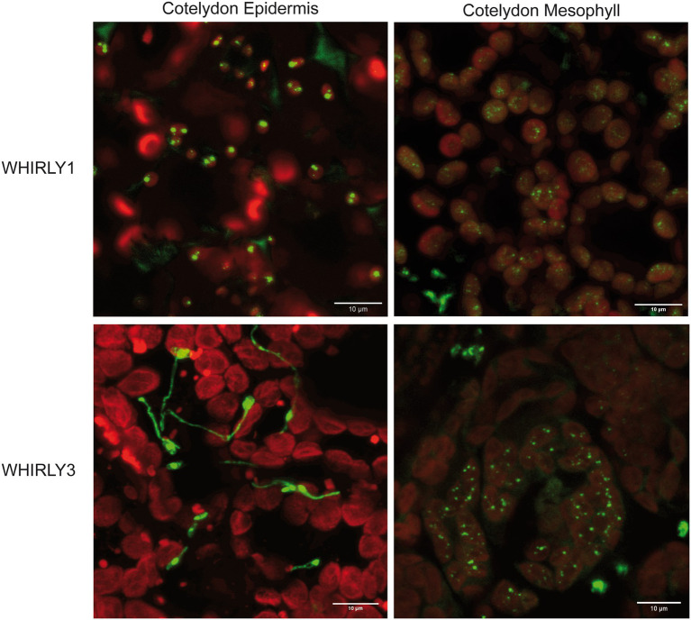
Figure 2.
Detection of WHIRLY1:GFP and WHIRLY3:RFP fusion proteins in epidermal cells of the cotyledons of stably transformed Arabidopsis plants. The constructs were overexpressed under the control of the CaMV 35S promoter. The constructs were overexpressed under the control of the CaMV 35S promoter. Leaves were imbedded in PBS:glycerol (1:1) without fixation and analyzed by a LEICA SP5 laser scanning microscope and a HCX PL Apo 63x/1.2 W objective. Sequential scans per frame for GFP [Ex 488 nm (6%), Em 510–550 nm], mRFP [Ex 543nm (14%), Em 580–610nm] and chlorophyll [Ex 633 (5%), Em 690–750 nm] were carried out. Chlorophyll fluorescence is shown in red while signals of WHIRLY1:GFP and WHIRLY3:RFP displayed both in green. A projection out of five optical layers representing 3 μm in z-direction are created by the LAS X software. The bars represent 10 μm each.

