
FIGURE 5.
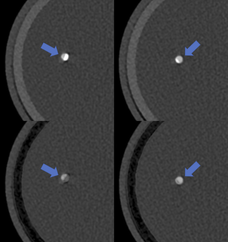
Axial example images at 65 keV of the 50% (left) and 25% (right) lesion for one of the vessels at a heart rate of 60 beats per minute. In comparison to the Mono reconstruction (top row), an improved visualization of the vessel lumen is depicted in the PureLumen reconstruction (bottom row), not affected by the potential overestimation from calcium blooming (lumen indicated by blue arrows).