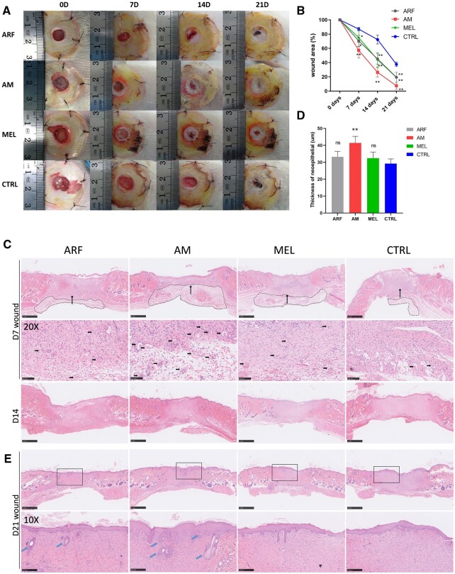
Figure 3.
ARF-MEL promotes chronic wound closure in STZ-induced diabetic rats. (A) Representative images depicting the process of wound closure on Days 0, 7, 14 and 21. (B) Wound closure rates were analyzed at each time point (one-way ANOVA). Data are presented as the means ± SD. n = 10. **P < 0.01 compared with the control group. Error bars represent SDs. (C) Representative H & E-stained images of wounds on Days 7 and 14, granulation tissue were indicated by the black dotted rectangular, scale bar, 1 mm; higher magnification images (×20) indicate an area denoted with long arrow in the upper image (Day 7), the small arrows point to the newly formed capillaries, scale bar, 100 μm. (D) Thickness of neoepithelial in wound edges was calculated in Day 7 (one-way ANOVA), n = 5 rats per group. **P < 0.01 compared with the control group. Error bars represent SDs. (E) Representative H & E-stained images of wounds on Day 21 and rectangles indicated the higher magnification observation area, scale bar, 1 mm; the arrows point to the hair follicles and sebaceous glands in higher magnification (×10), scale bar, 250 μm

