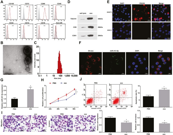
Figure 2.
hUCMSC-Exos suppress CRC cell growth. (A) Surface markers of hUCMSC-Exos were determined by flow cytometry; (B) hUCMSC-Exos were observed under a TEM; (C) particle diameter distribution of hUCMSC-Exos was analyzed by Zetasizer Nano ZS90; (D) protein expression of CD9, CD81, and TSG101 in hUCMSC-Exos was determined by western blot analysis; (E) uptake of hUCMSC-Exos by LoVo cells was observed under a fluorescent microscope; (F) the labeled fluorescent FITC-miR-431-5p and exosomes co-localization in LoVo cells was observed by the immunofluorescence microscopy, FITC-miR-431-5p labeled exosomes in green, 4ʹ,6-diamidino-2-phenylindole-stained nuclei were in blue; Dil-labeled exosomes were in red; (G) miR-431-5p expression in LoVo cells; (H) proliferation of LoVo cells was determined by MTT assay; (I) migration and invasion of LoVo cells were determined by Transwell assay; (J) apoptosis of LoVo cells was assessed using flow cytometry; *P < 0.05 versus the PBS group.

