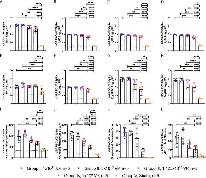
Fig 2. System serology profiling of anti-Spike antibodies from Ad26.COV2.S vaccinated NHPs.
(A) The humoral response against SARS-CoV-2 Spike was profiled using system serology. (B–G) The titer of IgG1 (B), IgG2 (C), IgG3 (D), IgG4 (E), IgM (F), and IgA (G) antibodies against SARS-CoV-2 were profiled using Luminex. (H, I) The titer of anti–SARS-COV-2 antibody binding to FcγRIIA-1 (H) and FcγRIII (I) were profiled using Luminex. (J–M) The graphs represent the ability of the humoral response to stimulate ADCP (J), neutrophil phagocytosis (ADNP) (K), complement deposition (ADCD) (L), or NK cell activation (NKdegran) (M) when stimulated with antigen-coated beads or plates. Bars represent the mean, and error bars represent the standard of deviation. This figure can be generated from the data found in data/figure2_data.csv of https://github.com/dzhu8/Ad26-Dose-Down. ADCD, antibody-dependent complement deposition; ADCP, antibody-dependent cellular phagocytosis; ADNP, antibody-dependent neutrophil phagocytosis; MFI, median fluorescent intensity; NHP, nonhuman primate; NK, natural killer; SARS-CoV-2, Severe Acute Respiratory Syndrome Coronavirus 2; VP, viral particle.

