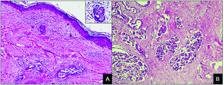
Fig 2. Photomicrographs of inflammatory mammary carcinoma in female dogs.
A) Proliferation of neoplastic cells in the superficial dermis associated with peritumoral inflammatory infiltrate. HE. Obj. 10x. Detail: Highly pleomorphic cells. HE. Obj. 40x. B) Proliferation of neoplastic cells invading the mammary parenchyma with developed stromal support. HE. Obj. 10x.

