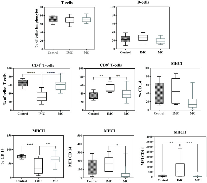
Fig 3. Leucocyte subsets in the peripheral blood of female dogs with mammary carcinoma.
Results are shown in box plot format, depicting frequencies of CD4+, CD8+, CD21+ T-lymphocytes, as well as CD14+CD45+, CD14+ MHC-I+ CD14+ MHC-II+ monocytes, and MFI MHCI and MFI MHC-II. Significant differences (P<0.05) are indicated by *, P<0.01 is indicated by **, P< 0.001 is indicated by *** and P< 0.0001 is indicated by ****.

