. 2019 Sep 11;9(49):28678–28687. doi: 10.1039/c9ra05725k

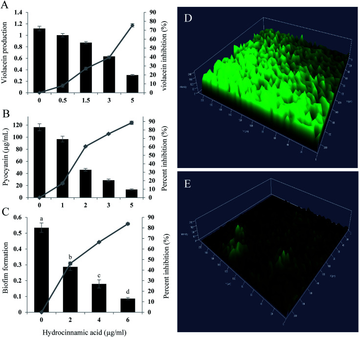
Fig. 4. QS inhibitory activity of hydrocinnamic acid. (A) C. violaceum CV026 grown in LB with various concentration of HCA for 24 h followed by extraction and quantification of violacein pigment. (B) Quantitative determination of pyocyanin inhibition in P. aeruginosa by hydrocinnamic acid. (C) Percentage of biofilm inhibition formed by P. aeruginosa represented by crystal violet staining for biomass. Confocal laser scanning micrographs of biofilm formed by P. aeruginosa untreated (D) and HCA treated (E). Data represent the mean ± SD of triplicate experiments. Different superscript letters indicate significant differences at P < 0.05.

Fig. 4