Figure 1.

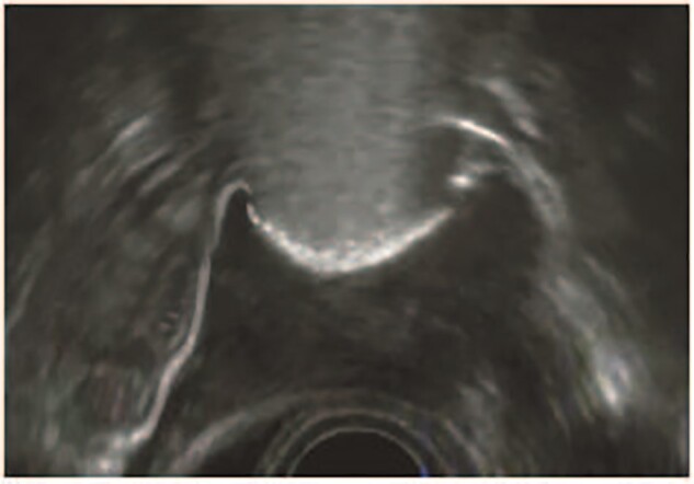
A typical 2D-hysterosalpingo-foam sonography image. The uterus is seen in transversal dimension with two patent fallopian tubes. Source: IQ Medical Ventures BV, Delft, the Netherlands.

A typical 2D-hysterosalpingo-foam sonography image. The uterus is seen in transversal dimension with two patent fallopian tubes. Source: IQ Medical Ventures BV, Delft, the Netherlands.