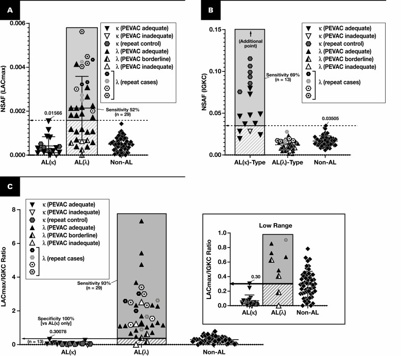
Figure 3.
Discrimination of AL(κ) and AL(λ). Results are plotted for cases of amyloidosis with at least triplicate analyses (scrapes) per slide. Each point represents the average of normalized spectral abundance factor (NSAF) results from scrapes with PEVAC (serum amyloid P, apolipoprotein E, victronectin, apoliprotein A4, and clusterin) of 2 or more. At the level of 100% specificity, the sensitivity for λ (A) and κ types (B) is marked. Cases that were repeated (hexagons, reflecting the control blocks) are counted only once in the sensitivity calculations. When replotting λ and κ light chain amyloidoses (AL(λ) and AL(κ), respectively) by the ratio of λ constant region to κ constant region (C), only two cases of putative AL(λ) type did not have a measured ratio greater than all cases of AL(κ), but neither were PEVAC adequate. LAC, immunoglobulin λ constant; LACmax, maximum of UniProt entries P0CG04, P0CG0, P0CG06, P0CF74, A0M8Q6.

