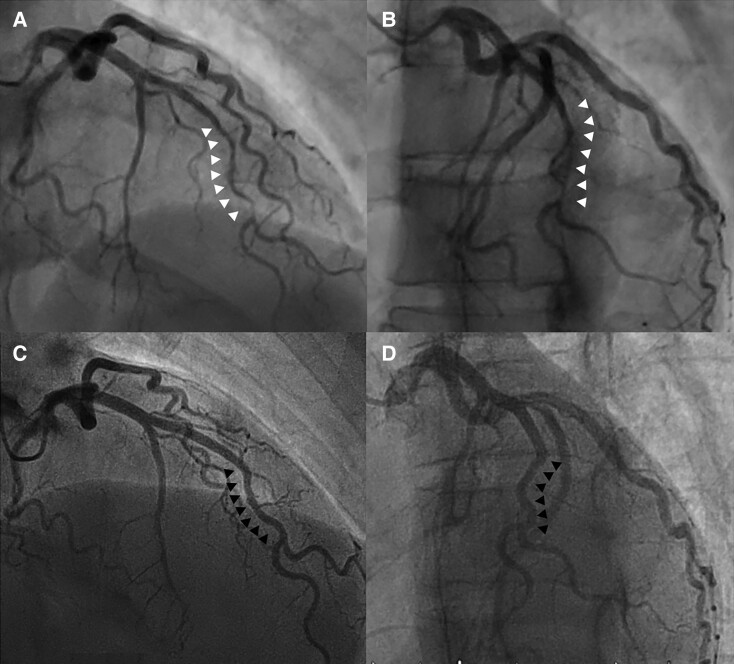
Figure 2.
(A and B) Coronary angiography in the right anterior oblique cranial view indicated Type 2A spontaneous coronary artery dissection based on the classification system published by Saw et al.9 involving the middle portion of the left anterior descending artery with a ‘stick insect’ appearance bordered by normal artery segment (white arrows) (C and D). Control angiography at 3M showed complete resolution of the previous pathological findings (black arrows). (A and C) Face cranial view. (B and D) Left anterior oblique cranial view.

